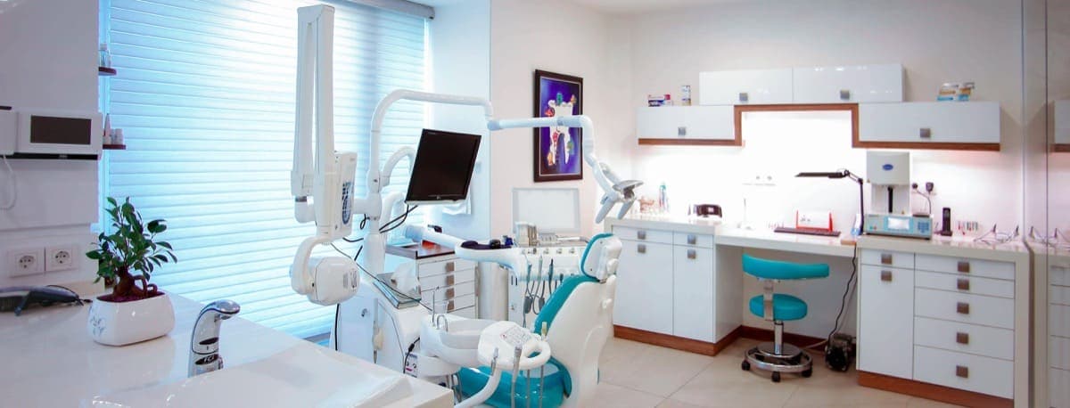
Expert Care for Your Vision
Your sight is precious. We provide advanced ophthalmology services to protect, preserve, and restore your vision with compassionate care.
Comprehensive Eye Care
Vision problems can be intimidating, but our team, led by Dr. Dibya Prabha, is here to help. We offer advanced diagnostics and personalized treatments for a wide range of eye conditions, prioritizing your comfort and understanding at every step. From routine care to complex retinal treatments, we are dedicated to your eye health. Our services include:
Targeted Retinal Therapy (Injections)
Administering medications directly into the retina to treat conditions like macular degeneration and diabetic retinopathy.
Key Benefits:
- Effective treatment for retinal diseases
- Minimally invasive procedure
- Quick recovery time
Laser Treatment for Retina
Using laser technology to treat retinal conditions such as diabetic retinopathy and retinal tears.
Key Benefits:
- Precise and targeted treatment
- Non-invasive procedure
- Prevents further vision loss
Advanced Surgery for Retinal Issues
Advanced surgical techniques to repair complex retinal conditions like retinal detachment and macular holes.
Key Benefits:
- High success rates
- Restores vision in severe cases
- Minimally invasive options available
Cataract Surgery
State-of-the-art cataract removal and lens replacement surgery to restore clear vision.
Key Benefits:
- Quick and painless procedure
- Improved vision within days
- Customized intraocular lenses (IOLs)
Detailed Retina Imaging (Fundus Photo)
High-resolution imaging of the retina to diagnose and monitor eye conditions.
Key Benefits:
- Non-invasive and painless
- Detailed images for accurate diagnosis
- Quick results for timely treatment
Advanced Eye Scans (OCT)
Non-invasive imaging for detailed retina and optic nerve analysis.
Key Benefits:
- High-resolution images
- Early detection of eye diseases
- Quick and painless procedure
Blood Flow Imaging (OCTA)
Advanced imaging to visualize blood flow in the retina and choroid.
Key Benefits:
- Non-invasive and detailed
- Early detection of vascular eye diseases
- Quick and painless procedure
Retinal Detachment Repair
Specialized care for retinal detachment, including surgical repair.
Key Benefits:
- High success rates
- Restores vision in severe cases
- Minimally invasive options available
Severe Eye Infection Treatment (Endophthalmitis)
Treatment for severe eye infections, including antibiotics and surgery.
Key Benefits:
- Prevents vision loss
- Customized treatment plans
- Quick and effective care
Macular Hole
Surgical repair of macular holes to restore central vision.
Key Benefits:
- High success rates
- Restores central vision
- Minimally invasive options available
Macular Degeneration Care (ARMD)
Comprehensive care for ARMD, including medications and laser therapy.
Key Benefits:
- Slows disease progression
- Preserves vision
- Personalized treatment plans
Eye Tumor Care (Ocular Oncology)
Specialized care for eye tumors, including diagnosis and treatment.
Key Benefits:
- Accurate diagnosis
- Customized treatment plans
- Preserves vision and eye health
Diagnostic Services
We offer a range of diagnostic services to accurately identify and monitor ophthalmological conditions. Our advanced technology ensures precise and timely diagnoses.
Optical Coherence Tomography (OCT)
Non-invasive imaging for detailed retina and optic nerve analysis.
Key Benefits:
- High-resolution images
- Early detection of eye diseases
- Quick and painless procedure
Visual Field Testing
Assessing peripheral vision to detect glaucoma and other eye conditions.
Key Benefits:
- Early detection of vision loss
- Customized treatment plans
- Non-invasive and quick
Common Conditions We Treat
Expert diagnosis and treatment for a wide range of eye conditions at Neurovision Clinic, Ranchi.
Cataract
A cataract is a clouding of the eye's natural crystalline lens that lies behind the iris and pupil. The lens works like a camera lens, focusing light onto the retina at the back of the eye. When it becomes cloudy, light cannot pass through clearly, resulting in blurred or dim vision — similar to looking through a fogged-up window. Cataracts develop slowly as part of the normal aging process (most people over 60 have some degree of cataract), but they can also develop due to diabetes, eye injury, prolonged steroid use, or excessive UV light exposure. Cataract surgery is the most commonly performed surgery worldwide and one of the safest and most effective surgical procedures, with over 98% success rate.
Glaucoma
Glaucoma is a group of eye diseases that damage the optic nerve — the vital cable that carries visual information from the eye to the brain. In most types of glaucoma, this damage is caused by abnormally high pressure inside the eye (intraocular pressure or IOP). However, glaucoma can also occur with normal eye pressure (normal-tension glaucoma). Over time, optic nerve damage leads to progressive, irreversible vision loss starting with peripheral (side) vision and eventually affecting central vision if untreated. Glaucoma is the second leading cause of blindness worldwide. Because early-stage glaucoma has no symptoms, regular comprehensive eye exams with optic nerve assessment are essential — especially for those with risk factors.
Diabetic Retinopathy
Diabetic retinopathy is a diabetes complication that affects the eyes. It is caused by damage to the blood vessels of the light-sensitive tissue at the back of the eye (retina). Chronically high blood sugar levels weaken and damage the tiny retinal blood vessels, causing them to leak fluid or bleed, distorting vision. In advanced stages, abnormal new blood vessels grow on the surface of the retina (proliferative diabetic retinopathy), which can lead to vitreous hemorrhage, retinal detachment, and severe vision loss. Diabetic retinopathy is the leading cause of blindness among working-age adults. The good news: with excellent blood sugar control, regular eye screening, and modern treatments available at Neurovision Clinic, over 90% of severe vision loss from diabetes can be prevented.
Retinal Detachment
Retinal detachment is a serious eye condition in which the retina — the light-sensitive layer of tissue at the back of the eye — separates from its underlying supportive tissue (the retinal pigment epithelium and choroid). Without its blood supply, the detached retina loses function, leading to vision loss. Retinal detachment is a medical emergency. There are three main types: rhegmatogenous (the most common, caused by a retinal tear or hole through which vitreous fluid seeps under the retina), tractional (scar tissue on the retina pulls it away, common in advanced diabetic retinopathy), and exudative (fluid accumulates under the retina without a tear, due to inflammation, tumors, or vascular conditions). Prompt surgical repair is essential to prevent permanent vision loss.
Age-Related Macular Degeneration (ARMD)
Age-Related Macular Degeneration (ARMD) is a degenerative eye disease that affects the macula — the small, central portion of the retina responsible for sharp, detailed central vision needed for reading, driving, recognizing faces, and seeing fine details. In ARMD, the macula gradually deteriorates. There are two forms: 'dry' (atrophic) ARMD, accounting for about 90% of cases, involves slow thinning of macular tissue and the buildup of drusen (yellow fatty deposits); 'wet' (neovascular or exudative) ARMD, while less common, is more aggressive — abnormal blood vessels grow beneath the macula (choroidal neovascularization), leaking fluid and blood, causing rapid and severe central vision loss. ARMD is the leading cause of severe vision loss in people over 50 in developed countries. It does NOT cause total blindness (peripheral vision is preserved) but profoundly affects quality of life.
Endophthalmitis
Endophthalmitis is a severe, sight-threatening infection involving the internal structures of the eye — the vitreous cavity (the gel filling the eye) and/or the aqueous humor (the fluid in the front of the eye). It is a true ophthalmic emergency. The infection can be introduced into the eye from outside (exogenous) — most commonly after eye surgery (postoperative), after an eye injection, or following penetrating eye trauma. Less commonly, it arrives via the bloodstream from an infection elsewhere in the body (endogenous). The most common causative organisms are bacteria (staphylococci, streptococci, gram-negative rods), though fungi can also cause endophthalmitis, especially in immunocompromised patients. Without rapid treatment, endophthalmitis leads to irreversible retinal damage, permanent vision loss, and potentially loss of the eye itself.
Macular Hole
A macular hole is a small break or defect that develops in the macula — the central, most sensitive part of the retina responsible for the sharp, detailed vision needed for reading, driving, and recognizing faces. Think of the macula as the 'HD center' of your vision. A macular hole creates a gap in this tissue, causing a blind spot or severe distortion in the center of your vision. Most macular holes are idiopathic (no specific cause), resulting from age-related changes in the vitreous gel that pull on the macula. They are most common in women over 60. Without treatment, macular holes typically enlarge over time and cause permanent central vision loss. Surgery (vitrectomy) is highly effective at closing the hole and improving vision.
Age-Related Macular Degeneration (ARMD)
Age-Related Macular Degeneration (ARMD or AMD) is the leading cause of severe, irreversible vision loss in people over 50 in developed countries. It affects the macula — the tiny, light-sensitive region at the center of the retina responsible for sharp central vision. In ARMD, the macula deteriorates over time. The 'dry' form (90% of cases) involves gradual thinning of macular tissue and accumulation of drusen deposits. The 'wet' form (10% of cases) is more aggressive — abnormal blood vessels grow beneath the macula, leaking fluid and blood, causing rapid vision loss. While peripheral vision is preserved (ARMD does not cause total blindness), the loss of central vision profoundly impacts reading, driving, recognizing faces, and independence. Modern treatments, especially anti-VEGF injections for wet ARMD available at Neurovision Clinic, have transformed outcomes — vision loss can now be halted or even reversed in many patients.
Ocular Tumors
Ocular oncology is the subspecialty of ophthalmology focused on the diagnosis and management of tumors and cancers affecting the eye and its surrounding structures. Ocular tumors can be benign or malignant (cancerous), primary (arising within the eye) or secondary (metastatic — spreading to the eye from another organ). The most common primary intraocular malignancy in adults is uveal (choroidal) melanoma. In children, retinoblastoma is the most common primary eye cancer. Other tumors include conjunctival melanoma, intraocular lymphoma, eyelid cancers (basal cell carcinoma, squamous cell carcinoma), and orbital tumors. Advances in imaging (OCT, OCTA, wide-field photography) now allow for highly accurate, non-invasive diagnosis and monitoring. Treatment has evolved from primarily removing the eye (enucleation) to eye-sparing approaches including radiation (plaque brachytherapy), laser, and in some cases, targeted molecular therapies.
Dry Eye Syndrome
Dry eye syndrome, also known as keratoconjunctivitis sicca, is a multifactorial disorder of the tear film and ocular surface resulting from either reduced tear production (aqueous-deficient) or excessive tear evaporation, most commonly due to meibomian gland dysfunction. A healthy tear film is essential for maintaining a smooth refractive surface, protecting against infection, and nourishing the avascular cornea. When tear film homeostasis is disrupted, the ocular surface becomes inflamed, leading to epithelial damage and neurosensory dysfunction. At Neurovision Clinic, Dr. Dibya Prabha provides comprehensive evaluation to identify the specific subtype, as treatment strategies differ significantly. Risk factors include advanced age, female gender, prolonged digital screen exposure, contact lens wear, refractive surgery, and certain systemic medications. In Ranchi's climate with seasonal dryness and dust, evaporative dry eye is particularly common. Dr. Prabha emphasizes that early intervention prevents chronic inflammation from causing irreversible damage to goblet cells and corneal nerves.
Refractive Error
Refractive error is the most common cause of correctable visual impairment worldwide, affecting approximately 2.3 billion people according to the WHO. It occurs when the optical power of the eye — determined by corneal curvature, lenticular power, and axial length — fails to focus light precisely on the fovea. The four classical categories are myopia (image focused anterior to the retina), hyperopia (posterior), astigmatism (meridional differences producing multiple focal points), and presbyopia (age-related loss of accommodative amplitude due to lenticular sclerosis). Uncorrected refractive error is the leading cause of avoidable visual impairment globally and the second most common cause of blindness. At Neurovision Clinic in Ranchi, Dr. Dibya Prabha provides comprehensive refraction services using subjective and objective techniques including autorefraction, retinoscopy, and cycloplegic refraction in children. She emphasizes that proper refractive correction improves visual acuity, quality of life, academic performance in children, and workplace safety.
Conjunctivitis
Conjunctivitis, commonly known as pink eye, is inflammation of the conjunctiva — the thin transparent mucous membrane lining the inner eyelids and covering the sclera up to the corneal limbus. It is the most common cause of acute red eye worldwide, classified into infectious (viral, bacterial, chlamydial) and non-infectious (allergic, irritant, medication-induced) types. Viral conjunctivitis, predominantly adenoviral, accounts for up to 80% of acute cases. Bacterial conjunctivitis is more common in children, with Haemophilus influenzae, Streptococcus pneumoniae, and Moraxella catarrhalis as leading isolates. Allergic conjunctivitis affects 15–20% of the population and is frequently associated with rhinitis and atopic dermatitis. At Neurovision Clinic in Ranchi, Dr. Dibya Prabha provides rapid etiological diagnosis and targeted therapy, recognizing that inappropriate use of antibiotics or corticosteroids can prolong infection or cause steroid-induced glaucoma. She emphasizes that any red eye with pain, photophobia, or decreased vision warrants urgent evaluation to exclude keratitis, anterior uveitis, and acute angle-closure glaucoma.
Keratoconus
Keratoconus is a progressive, bilateral (though often asymmetric) corneal ectatic disorder in which the cornea thins and assumes a conical shape, resulting in irregular astigmatism and visual distortion. The pathogenesis involves genetic predisposition, biomechanical corneal instability, and enzymatic degradation of stromal collagen by matrix metalloproteinases with reduced tissue inhibitors (TIMPs). Histologically, it features fragmentation of Bowman's layer, stromal thinning, and iron deposition in the basal epithelium (Fleischer ring). Fine vertical striae in the deep stroma (Vogt's striae) are pathognomonic, while acute hydrops occurs when Descemet's membrane ruptures, allowing aqueous to enter the stroma. The condition typically presents in the second decade of life and may progress for 10–20 years before stabilizing. At Neurovision Clinic in Ranchi, Dr. Dibya Prabha provides comprehensive keratoconus care from early detection through corneal topography to advanced management. She educates patients that eye rubbing is the strongest modifiable risk factor, and that atopic conditions such as vernal keratoconjunctivitis increase susceptibility.
Uveitis (Intraocular Inflammation)
Uveitis is an inflammatory condition affecting the uveal tract, the middle vascular layer of the eye consisting of the iris, ciliary body, and choroid. It can present acutely or chronically and may involve one or both eyes. The inflammation arises when the body's immune system attacks ocular tissues, triggered by infection, systemic autoimmune disease, or occasionally trauma. Uveitis is categorised anatomically: anterior uveitis (iritis) is the most common form, presenting with a red, painful eye; intermediate uveitis involves the vitreous cavity; posterior uveitis affects the retina and choroid; and panuveitis involves all layers. In India, infectious causes such as tuberculosis and toxoplasmosis are significant contributors. Uveitis can occur at any age but is most prevalent in adults between 20 and 60 years. If inadequately treated, it is a leading cause of preventable blindness, responsible for up to 10% of visual impairment cases worldwide.
Squint (Strabismus)
Squint, known medically as strabismus, is an ocular misalignment where the visual axes of the two eyes are not simultaneously directed at the same object of regard. The condition can be manifest (tropia), where the deviation is constantly present under binocular viewing conditions, or latent (phoria), where the misalignment is controlled by fusion mechanisms and appears only when fusion is disrupted. Squint is classified by direction: esotropia is inward deviation, exotropia outward, hypertropia upward, and hypotropia downward. Based on constancy, it may be intermittent or constant, and by laterality, unilateral or alternating. Squint affects approximately 2-4% of the population and can present at any age. In children, the most critical consequence is the development of amblyopia, as the brain suppresses the image from the deviating eye. In adults, acquired strabismus frequently causes diplopia and impaired depth perception, significantly affecting daily activities and quality of life.
Corneal Ulcer (Infectious Keratitis)
A corneal ulcer, or infectious keratitis, is a breach in the corneal epithelium with underlying stromal infiltration, suppuration, and tissue necrosis caused by microbial invasion. It represents one of the most common preventable causes of corneal blindness worldwide, with an estimated 1.5 to 2 million new cases annually, predominantly in low- and middle-income countries. The causative organisms include bacteria (Staphylococcus aureus, Streptococcus pneumoniae, Pseudomonas aeruginosa), fungi (Fusarium, Aspergillus, Candida), viruses (herpes simplex, herpes zoster), and protozoa (Acanthamoeba). In India, particularly in agrarian states like Jharkhand, fungal keratitis accounts for a significant proportion of corneal ulcers due to corneal trauma with vegetative matter. Bacterial keratitis tends to be more acute and aggressive, with rapid stromal melting, while fungal keratitis follows a subacute course with feathery, dry infiltrates. The presence of a hypopyon, a layer of inflammatory cells in the anterior chamber, indicates severe disease regardless of the aetiology. Without timely and appropriate treatment, corneal ulcers can result in permanent visual impairment from corneal scarring, irregular astigmatism, thinning, descemetocele, or frank perforation.
Pterygium (Nakhuna)
Pterygium, known as nakhuna in Hindi due to its resemblance to a fingernail growing over the eye, is a benign fibrovascular proliferation of conjunctival tissue that encroaches onto the cornea in a triangular or wing-shaped configuration. The lesion consists of a head (the apical portion on the cornea), a neck, and a body (the bulbar conjunctival portion). Histologically, it demonstrates elastotic degeneration of subepithelial collagen, believed to result from chronic actinic (ultraviolet) damage. Pterygium is strongly associated with cumulative UV-B exposure and is therefore more prevalent in tropical and subtropical regions, with prevalence rates in some Indian populations exceeding 10%. It is typically located on the nasal limbus, as the cornea's nasal aspect receives focused UV radiation reflected and refracted from the nasal bridge. Pterygium occurs bilaterally in many patients, though often asymmetrically. While classified as a benign lesion, progressive pterygia can cause significant visual morbidity through direct corneal invasion, induced astigmatism, chronic ocular surface inflammation, and tear film instability. At Neurovision Clinic, Ranchi, Dr. Dibya Prabha provides comprehensive pterygium care ranging from conservative management to advanced surgical excision with conjunctival autograft.
Advanced Diagnostic Tests
We use state-of-the-art technology to accurately diagnose and monitor eye conditions.
OCT (Optical Coherence Tomography)
Optical Coherence Tomography (OCT) is a advanced, non-invasive imaging test that uses light waves to create high-resolution, cross-sectional images of the retina — the light-sensitive tissue lining the back of the eye. Think of it as an 'optical ultrasound' that can visualize the retina's distinct layers with incredible detail, down to a few microns (thousandths of a millimeter). OCT can detect subtle retinal changes long before they cause visible symptoms, making it invaluable for early diagnosis and monitoring of conditions like diabetic retinopathy, macular degeneration, glaucoma, and macular holes. At Neurovision Clinic, Dr. Dibya Prabha uses OCT as a cornerstone of retinal evaluation — the test takes only a few minutes and provides immediate, detailed information about your retinal health.
OCTA (OCT Angiography)
OCTA (Optical Coherence Tomography Angiography) is the latest advancement in retinal imaging that captures detailed 3D maps of blood vessels in the retina and choroid — entirely without injecting any dye. It uses the movement of red blood cells through retinal vessels to create high-resolution images of blood flow. Compared to traditional fluorescein angiography which requires IV dye injection and takes 20+ minutes, OCTA is completely non-invasive, takes only seconds, and provides depth-resolved images showing exactly which retinal layer is affected. At Neurovision Clinic, Dr. Dibya Prabha uses OCTA to detect and monitor retinal vascular diseases with unprecedented precision.
Fundus Photography
Fundus photography is a specialized ophthalmic imaging technique that captures high-resolution, full-color photographs of the fundus — the interior back surface of the eye including the retina, optic nerve head, macula, and retinal blood vessels. A fundus camera is essentially a specialized low-power microscope with an attached camera. The images serve as a permanent, objective record of retinal appearance, allowing Dr. Dibya Prabha at Neurovision Clinic to precisely monitor even subtle changes over time. This is critically important for chronic, progressive conditions like diabetic retinopathy, glaucoma, and age-related macular degeneration where year-over-year comparison guides treatment decisions.
Visual Field Testing (Perimetry)
Visual field testing, also called perimetry, is a systematic method of measuring the entire scope of a person's vision — both central detail vision and peripheral (side) awareness. The test maps the sensitivity of vision across the visual field, detecting areas of reduced or lost vision (scotomas) that the patient may not be consciously aware of. At Neurovision Clinic, Dr. Dibya Prabha uses automated perimetry to evaluate and monitor a range of conditions: Diagnose and monitor glaucoma — peripheral vision loss is often the first sign Assess optic nerve damage from neurological conditions including optic neuritis and ischemic optic neuropathy Evaluate vision loss after stroke affecting visual pathways in the brain Monitor for hydroxychloroquine (Plaquenil) retinal toxicity in rheumatology patients Evaluate unexplained vision loss — differentiate retinal from neurological causes
Retinal Laser Treatment (Laser Photocoagulation)
Retinal laser treatment, medically known as laser photocoagulation, is a therapeutic procedure that uses a precisely focused beam of high-energy light to treat specific retinal conditions. The laser creates microscopic, controlled thermal burns on the retina, which seal leaking blood vessels, create a barrier around retinal tears to prevent retinal detachment, or reduce abnormal blood vessel growth (neovascularization). Different types of retinal laser are used depending on the condition being treated: Focal/grid laser for diabetic macular edema — seals leaking microaneurysms in the macula Pan-retinal photocoagulation (PRP) for proliferative diabetic retinopathy — reduces abnormal blood vessel growth across the retina Laser retinopexy for retinal tears — creates a barrier of scar tissue around the tear to prevent retinal detachment Laser for retinopathy of prematurity — treats abnormal blood vessels in premature infants Dr. Dibya Prabha, with her Retina & Vitreous fellowship training from LV Prasad Eye Institute Hyderabad, performs retinal laser treatments with precision and clinical expertise at Neurovision Clinic, Ranchi.
LASIK (Laser-Assisted In Situ Keratomileusis)
LASIK (Laser-Assisted In Situ Keratomileusis) is the most commonly performed laser eye surgery worldwide for correcting refractive errors — nearsightedness (myopia), farsightedness (hyperopia), and astigmatism. The procedure uses a precise excimer laser to reshape the cornea, the clear front surface of the eye, so that light entering the eye focuses correctly on the retina. During the procedure, a thin flap is created on the corneal surface using a femtosecond laser (bladeless LASIK), the flap is gently lifted, the underlying corneal tissue is reshaped with the excimer laser, and the flap is repositioned where it heals naturally without stitches. The entire procedure takes about 15 minutes for both eyes. At Neurovision Clinic in Ranchi, Dr. Dibya Prabha (MS Ophthalmology) evaluates each patient thoroughly — including corneal topography, pachymetry (corneal thickness measurement), tear film assessment, and refraction — to determine LASIK candidacy. Only patients who meet strict safety criteria are offered the procedure. For patients who are not ideal LASIK candidates, alternative vision correction options including PRK, ICL (Implantable Collamer Lens), and refractive lens exchange are discussed.
Vitrectomy (Vitreoretinal Surgery)
Vitrectomy is a microsurgical procedure in which the vitreous gel — the clear, jelly-like substance filling the center of the eye — is removed to allow the surgeon direct access to the retina and other posterior segment structures. The vitreous is replaced with a balanced salt solution, gas bubble, or silicone oil depending on the condition being treated. Modern vitrectomy uses small-gauge instruments (23G, 25G, or 27G) that pass through tiny, self-sealing incisions in the sclera, typically not requiring sutures. Vitrectomy is the foundational procedure for treating a wide range of retinal conditions: retinal detachment repair, macular hole closure, epiretinal membrane peeling, removal of vitreous hemorrhage, management of advanced diabetic retinopathy, retrieval of intraocular foreign bodies, and complications of cataract surgery. At Neurovision Clinic in Ranchi, Dr. Dibya Prabha (MS Ophthalmology) evaluates each patient with comprehensive retinal imaging — including OCT (Optical Coherence Tomography), OCTA (OCT Angiography), fundus photography, and B-scan ultrasonography when the view is obscured — before recommending vitrectomy. The procedure is performed in a fully equipped operating theatre with strict aseptic protocols.
Corneal Transplant (Keratoplasty)
Corneal transplant, or keratoplasty, is a microsurgical procedure in which damaged or diseased corneal tissue is removed and replaced with healthy donor corneal tissue obtained through eye donation. The cornea is the transparent front window of the eye — when it becomes scarred, irregular, or opaque from disease or injury, vision is severely impaired. Corneal transplant types include: penetrating keratoplasty (PK — full-thickness replacement of all five corneal layers), deep anterior lamellar keratoplasty (DALK — replacement of the front layers while preserving the patient's own healthy endothelial layer, reducing rejection risk), and Descemet stripping endothelial keratoplasty (DSEK/DMEK — selective replacement of only the innermost endothelial layer through a small incision, with faster recovery). At Neurovision Clinic in Ranchi, Dr. Dibya Prabha evaluates each patient with corneal topography, pachymetry, slit-lamp examination, and endothelial cell analysis to determine which procedure offers the best balance of visual outcome and safety. The clinic coordinates with eye banks for timely access to high-quality donor corneal tissue. Corneal transplant is one of the most successful forms of human tissue transplantation, with graft survival rates exceeding 90% in the first year for low-risk cases.
Pediatric Eye Surgery
Pediatric eye surgery encompasses a range of surgical procedures performed on infants, children, and adolescents to correct eye conditions that threaten vision development, ocular alignment, or eye health. Children's eyes are not simply smaller versions of adult eyes — the developing visual system imposes unique urgency, as conditions like congenital cataract, uncorrected refractive errors, and strabismus (misaligned eyes) can cause amblyopia (lazy eye) if not treated during the critical period of visual development (birth to approximately 8–10 years). Pediatric eye surgeries at Neurovision Clinic, Ranchi include: strabismus surgery (squint correction) to realign the eye muscles, congenital cataract surgery with or without intraocular lens implantation, probing and syringing for congenital nasolacrimal duct obstruction (blocked tear duct), ptosis surgery (drooping eyelid repair) when the eyelid obstructs vision, and examination under anaesthesia (EUA) for children who cannot cooperate with in-clinic examination. Dr. Dibya Prabha (MS Ophthalmology) has extensive experience managing pediatric eye conditions and works closely with parents to ensure the surgical plan, anaesthesia safety, and post-operative care are clearly understood. All pediatric procedures are performed in a facility with paediatric anaesthesia capability.
Oculoplasty (Oculoplastic Surgery)
Oculoplasty, also known as oculoplastic surgery or ophthalmic plastic and reconstructive surgery, is a subspecialty of ophthalmology focused on the eyelids, tear drainage system (lacrimal system), orbit (the bony socket surrounding the eye), and the surrounding facial structures. Oculoplastic procedures address both functional problems (drooping eyelids obstructing vision, eyelids turning inward or outward, blocked tear ducts causing chronic watering) and reconstructive or cosmetic concerns (eyelid bag removal, repair of eyelid defects after tumour excision, reconstruction after trauma). Common oculoplastic procedures at Neurovision Clinic, Ranchi include: ptosis repair (tightening the levator muscle or performing a frontalis sling for severe ptosis), DCR (dacryocystorhinostomy — creating a new tear drainage passage for blocked nasolacrimal ducts), entropion and ectropion repair (correcting inward or outward turning eyelids), blepharoplasty (removal of excess eyelid skin and fat), eyelid tumour excision with reconstruction, eyelid laceration repair, and enucleation or evisceration (removal of a blind, painful eye) with orbital implant placement. Dr. Dibya Prabha (MS Ophthalmology) evaluates each patient with a thorough eyelid, lacrimal, and orbital examination to determine the precise surgical approach. Most oculoplastic procedures are performed under local anaesthesia with sedation, allowing same-day discharge.
Squint Surgery (Strabismus Surgery)
Squint surgery, also called strabismus surgery or eye muscle surgery, is a procedure that corrects misalignment of the eyes by adjusting the position or tension of the extraocular muscles — the six muscles attached to the outside of each eye that control all eye movements. When one or more of these muscles is too strong or too weak, the eyes do not point in the same direction, causing squint (strabismus). The misalignment may be constant or intermittent, and may affect one eye or alternate between both eyes. Squint surgery involves weakening an overacting muscle (by recessing it — detaching and reattaching it further back on the sclera) or strengthening an underacting muscle (by resecting it — removing a segment and reattaching the shortened muscle). Adjustable suture techniques allow fine-tuning of the alignment in the immediate post-operative period for cooperative adults. Squint surgery addresses both cosmetic concerns (restoring straight eyes and a normal appearance) and functional concerns (preserving or recovering binocular vision, depth perception, and preventing amblyopia in children). At Neurovision Clinic in Ranchi, Dr. Dibya Prabha (MS Ophthalmology) performs a complete orthoptic evaluation — including cover tests, prism measurements, ocular motility assessment, and stereopsis testing — before recommending surgery. For children, surgery timing is coordinated around the critical period of visual development. For adults, squint surgery can restore eye alignment and improve quality of life at any age.
Common Eye Symptoms
Expert evaluation and treatment for eye-related symptoms at Neurovision Clinic, Ranchi.
Vision Loss
Sudden or gradual vision loss is a medical emergency. Dr. Dibya Prabha, MS Ophthalmology (RIMS), FICO, Retina Fellow (LV Prasad Eye Institute, Hyderabad), provides expert evaluation and treatment at Neurovision Clinic, Ranchi.
Eye Pain
Eye pain is your body's warning signal. Dr. Dibya Prabha, MS Ophthalmology (RIMS), FICO, Retina Fellow, provides precise diagnosis and effective treatment for all causes of eye pain at Neurovision Clinic, Ranchi.
Double Vision (Diplopia)
Double vision is disorienting, disabling, and often a sign of a serious underlying condition. Dr. Dibya Prabha (MS Ophthalmology, FICO, Retina Fellow LV Prasad Eye Institute Hyderabad) provides expert evaluation to determine whether diplopia originates from the eye or the nervous system, and offers treatment — from prisms to coordinated neurological referral — at Neurovision Clinic, Ranchi.
Eye Redness
A red eye may be as simple as a subconjunctival hemorrhage or as serious as a vision-threatening uveitis or keratitis. Dr. Dibya Prabha (MS Ophthalmology, FICO, Retina Fellow LV Prasad Eye Institute Hyderabad) provides expert slit lamp examination to diagnose the cause of eye redness and deliver targeted treatment at Neurovision Clinic, Ranchi.
Flashes & Floaters
New onset of flashes and floaters is a warning signal from your retina. Dr. Dibya Prabha (MS Ophthalmology, FICO, Retina Fellow LV Prasad Eye Institute Hyderabad) provides same-day urgent evaluation to rule out retinal tears and detachment at Neurovision Clinic, Ranchi.
Night Blindness (Nyctalopia)
Struggling to see in dim light or at night? Night blindness (nyctalopia) can signal an underlying retinal, nutritional, or ocular condition. Dr. Dibya Prabha (MS Ophthalmology, FICO, Retina Fellow LV Prasad Eye Institute Hyderabad) provides expert diagnosis and treatment at Neurovision Clinic, Ranchi — because safe mobility after dark should not be a privilege.
Dry Eyes
Chronic dry eye is more than discomfort — it is an inflammatory disease of the ocular surface that can damage the cornea and degrade vision. Dr. Dibya Prabha (MS Ophthalmology, FICO, Retina Fellow LV Prasad Eye Institute Hyderabad) provides comprehensive dry eye evaluation and personalized, evidence-based treatment at Neurovision Clinic, Ranchi.
Watery Eyes (Epiphora)
Constantly watery eyes are not just a social nuisance — they can signal an underlying tear drainage obstruction, ocular surface disease, or eyelid disorder. Dr. Dibya Prabha (MS Ophthalmology, FICO, Retina Fellow LV Prasad Eye Institute Hyderabad) provides expert diagnosis and treatment at Neurovision Clinic, Ranchi.
Itchy Eyes (Ocular Pruritus)
Persistent eye itching is the hallmark of ocular allergy — but it can also signal blepharitis, dry eye, or contact dermatitis. Dr. Dibya Prabha (MS Ophthalmology, FICO, Retina Fellow LV Prasad Eye Institute Hyderabad) provides expert diagnosis and targeted treatment at Neurovision Clinic, Ranchi — so you can stop rubbing and start living comfortably.
Blurred Vision
Blurred vision — whether gradual or sudden, in one eye or both — always demands a thorough eye examination. Dr. Dibya Prabha (MS Ophthalmology, FICO, Retina Fellow LV Prasad Eye Institute Hyderabad) provides meticulous, systematic evaluation to pinpoint the cause and restore clarity at Neurovision Clinic, Ranchi.

Book Your Visit Today
Get expert care for brain, eye, and spine conditions in Ranchi. Serving Jharkhand, Bihar, West Bengal, and Chhattisgarh. Book now on WhatsApp—we're here to help.